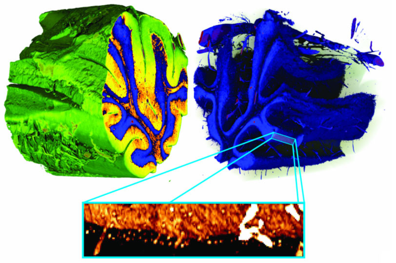
Three-dimensional false-colour renderings of a human cerebellum, obtained with X-ray grating interferometric phase-contrast tomography.
Figure 1: Three-dimensional false-colour renderings of a human cerebellum, obtained with X-ray grating interferometric phase-contrast tomography. White matter (orange) can be distinguished from two types of grey matter (blue: stratum granulosum; yellow: stratum moleculare). The data also show blood vessels and individual cells identified as Purkinje cells (detail inset at bottom of figure).